
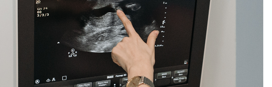
1. Definition
Endoscopic Ultrasound (EUS) is a minimally invasive diagnostic and therapeutic procedure that combines:
2. Purpose
3. When is it recommended?

Primary type used to diagnose conditions.
Evaluates the walls of the GI tract, pancreas, bile ducts, and surrounding structures.
Identifies tumors, cysts, and inflammation.
Combines EUS with needle biopsy.
Used to obtain tissue samples from suspicious areas.
Helps diagnose cancer, infections, or inflammation.
Similar to FNA but uses a different needle design.
Provides larger tissue samples.
More effective for diagnosing pancreatic and GI tumors.
Combines imaging with minimally invasive treatments.
Used for:
These procedures are recommended for patients experiencing:
Chronic abdominal pain.
Unexplained weight loss.
Persistent nausea or vomiting.
Blood in stool or black, tarry stools.
Vomiting blood (hematemesis).
Difficulty swallowing (dysphagia).
Esophageal strictures or tumors.
Yellowing of the skin and eyes.
Suggests bile duct obstruction or liver problems.
Chronic diarrhea or weight loss.
Suspected pancreatic or bile duct disease.
Esophageal, pancreatic, stomach, and bile duct cancers.
EUS detects and stages the tumors.
Inflammation of the pancreas.
EUS visualizes structural changes.
Gallstones or bile duct strictures.
EUS locates and assesses blockages.
Pancreatic cysts or pseudocysts.
EUS helps in diagnosis and drainage.
Swollen lymph nodes due to cancer or infection.
EUS aids in biopsy and staging.
Balanced diet with fiber, fruits, and vegetables.
Avoid processed and fatty foods.
Stay hydrated.
Reduces the risk of GI cancers and pancreatitis.
Promotes digestive health.
Routine check-ups for at-risk individuals.
Early detection of GI issues reduces the need for invasive procedures.
Use PPIs or H2 blockers to reduce acid reflux.
Prevents esophageal damage.
Healthy weight lowers the risk of gallstones and GI issues.
Reduces inflammation in the digestive system.
PPIs and H2 blockers for GERD.
Antibiotics for infections.
Pain management for chronic conditions.
Used to drain pancreatic pseudocysts or abscesses.
Minimally invasive alternative to surgery.
Fine-needle aspiration (FNA) for tissue sampling.
Determines cancer stage and spread.
EUS-guided celiac plexus neurolysis for chronic pain.
Biliary and pancreatic drainage procedures.
Fasting for 6-8 hours before the procedure.
Medication adjustments (if required).
Sedation or anesthesia is administered.
Patient lies on their side.
Endoscope with ultrasound probe inserted through the mouth or rectum.
Ultrasound waves create detailed images of internal organs.
Fine-needle aspiration (FNA) or biopsy may be performed.
Therapeutic procedures (e.g., drainage) may be carried out if needed.
Mild throat discomfort for a few hours.
Rest and hydration are recommended.
Avoid eating or drinking until swallowing returns to normal.
Continue prescribed medications if applicable.
Avoid NSAIDs or blood thinners temporarily.
Results are reviewed with the patient.
Follow-up imaging or biopsy results discussed.


